(509) 228-1000 WA
(208) 754-3100 ID

PET/CT

Share

CCNW offers PET/CT scans, a safe, painless, non-invasive procedure. It is the combination of both a PET scan and a CT scan


What is a PET/CT?

A PET/CT scan is a safe, painless, non-invasive procedure. It is the combination of both a PET scan and a CT scan. It is one continuous body scan in which the functions of a PET scan catch the small changes in the body’s metabolism due to the abnormal growth of cells. The CT function of the machine helps physicians find the exact size, location and shape of the tumor or disease in question.

By integrating the PET and CT technologies it makes it possible to get both the anatomical and biological data of the tumor in just one exam. This approach makes it easier to detect various cancers including breast, melanoma, lymphoma, lung and more. In addition, it simplifies the treatment plan including type of surgical procedure, response and effectiveness to treatments and the overall extent of the disease.


Why should I get a PET/CT?

There are many benefits of having a PET/CT including earlier detection of the cancer, followed by accurate staging and location of the tumor ending with detailed treatment and monitoring.

In the past, many complications arose from trying to interpret the PET and CT scans separately due to changes in the body between scans. Additionally, unnecessary procedures, misdiagnosis and longer treatment resulted with the separate scans. With the PET/CT, many of the past situations can now be avoided.


How does it work?

To begin the procedure, a small amount of radioactive glucose, “FTG,” is injected into your bloodstream. There is no danger to you from this injection. Glucose (also known as sugar) is a common substance every cell in your body needs in order to function. The PET/CT exposes you to a very low level of radiation.

After the injection, you will wait approximately an hour while the injection material is distributed throughout your body. Then you will be asked to lie on a table that passes slowly through the scanner. 

When disease occurs, the biochemistry of your tissues and cells changes. In cancer, for example, cells begin to grow at a much faster rate and metabolize (break down) sugar at a higher rate than normal cells. If an area in an organ is cancerous, the signals will be stronger than in the normal surrounding tissue. A scanner records this data and transforms it into pictures. Abnormal cells or tissue will appear as bright spots. Normal, healthy organs and tissues will not have the bright glow that abnormal cells exhibit in a PET/CT scan.

 
History of PET/CT

Before the PET/CT, doctors would have to run both a PET and a CT scan and then attempt to match them in order to determine the approximate location and size of the tumor. They took the separate images and made their “best guess” as to gaining information about the tumor. Then, in 1992, engineer Ron Nutt and physicist David Townsend created the idea to combine the two machines into one.

Nutt and Townsend worked on this concept for three years before receiving any additional assistance until they received a grant from the National Cancer Institute.  Their idea was that the machine should be more patient-friendly so they created the machines to be 28 inches in diameter for the tunnel.  The machine was finally completed in 1998 and installed at the University of Pittsburg medical center.


PET/CT at Cancer Care Northwest

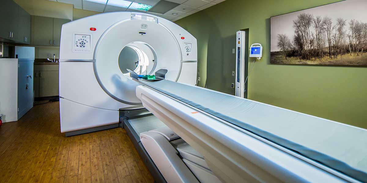
Cancer Care Northwest is home to the GE Discovery IQ 4-Ring PET/CT Scanner, the most sensitive imaging technology available today. Our PET/CT scanner is the busiest scanner in the Inland Northwest and is located within our South Spokane treatment center (601 S Sherman Street).

This sophisticated scanner not only produces pictures that show the location and nature of the tumor, but it also takes four-dimensional (4D) images that capture a tumor in motion. For example, a lung tumor moves as a patient is breathing. With this 4D information, the radiation team can precisely pinpoint a tumor’s location at any given moment, allowing your doctors to develop a more effective treatment plan.

Cancer Care Northwest is home to the GE Discovery IQ PET/CT Scanner, the most sensitive imaging technology available today.


Molecular Imaging - Gallium-68 (GA-68) Dotatate PET/CT scans

Cancer Care Northwest offers Gallium-68 (GA-68) Dotatate PET/CT scans for the localization of neuroendocrine tumors (NETs) that are somatostatin receptor positive. NET tumors develop most commonly in the lungs, appendix, small intestine, rectum, and pancreas and produce an “overexpression” of a specific cell feature called somatostatin receptors. Molecular imaging technologies use this cell feature to detect cancerous cells throughout the body and as a target for the delivery of therapy. A type of molecular imaging, GA-68 PET/CT scans, may be performed during initial (diagnostic) testing to locate a cancer tumor or to look at where the NET has spread. It may also be done at other times to check how the treatment is working or if tumor cells have spread, or metastasized.

Performed at the CCNW South Clinic (601 S Sherman St), patients can expect a GA-68 PET/CT scan to take approximately 2 hours. The GA-68 doctorate “radioactive tracer” will be given intravenously to the patient. Cells with somatostatin receptors on their surface will attract and attach to the Ga-68 dotatate. You will then have a PET scan, which can detect the radiation put off by the Ga-68 dotatate. The scanner's computer creates a figure of the patient on the screen. Any areas with a higher amount of GA-68 will show up as a bright spot on the image.

The benefits to using GA-68 PET/CT scans include improved imaging accuracy and increased imaging sensitivity and specificity, which help oncologists to diagnose, stage and treat the cancer, and in disease management. Additionally, molecular imaging in patients with NET can help with localization of disease (primary) and to evaluate the extent of the disease.


Axumin® PET/CT Scans for Recurrent Prostate Cancer
CCNW is proud to be the only healthcare facility in Eastern Washington to currently offer Axumin PET/CT scans for patients with recurrent prostate cancer. Axumin® (fluciclovine F 18) is an advanced diagnostic imaging agent, or radiotracer, that is used in conjunction with a positron emission tomography (PET)/computed tomography (CT) scan for men who have had prior treatment for prostate cancer and now have elevated Prostate Specific Antigen (PSA) levels.
 
Approved by the FDA in 2016, Axumin is a Medicare-covered scan that can locate recurrent prostate cancer after surgery or radiation in patients with a PSA level less than 10. In years past, bone and body scans have been unable to determine the location and extent of the cancer until the PSA level is excessively elevated (10 – 30 or higher). The Axumin PET scan works by detecting the metabolic activity of the cancer itself, allowing clinicians to detect small local (prostate bed) and distant (lymph node, bone) metastatic lesions earlier than ever before when PSA levels are less than 10. 
 
Performed at the CCNW South Clinic (601 S Sherman St), patients can expect an Axumin PET/CT scan to take approximately 30 minutes. The Axumin “radioactive tracer” will be given intravenously to the patient 3-5 minutes prior to the scan. The Axumin links to an amino acid, which is absorbed by prostate cancer at a much more rapid rate than normal cells. The rapid absorption of Axumin by prostate cancer cells is then imaged using PET scan equipment. Location of high areas of radiation signal the location of the cancer in the patient’s body.
 
Full Axumin prescribing information is available at www.axumin.com. Access the Axumin Patient Brochure here.
 
 
GA-PSMA-11 Injection PET/CT for Prostate Cancer

Cancer Care Northwest now offers advanced molecular imaging service, Gallium Ga-68 gozetotide injection, also known as Ga-PSMA-11 injection. CCNW is one of the first healthcare providers in the Spokane area to offer this new and highly specific PSMA PET imaging agent that can help health care professionals diagnose the stage and spread of disease – an important step for the optimal care of men with prostate cancer.

The Ga-PSMA-11 injection is indicated for positron emission tomography (PET) of prostate specific membrane antigen (PSMA) positive lesions in patients with prostate cancer with:

  • suspected metastasis who are candidates for initial definitive therapy;
  • suspected recurrence based on elevated serum prostate-specific antigen (PSA) level.

Prostate cancer is the most common cancer in American men after skin cancer. According to the American Cancer Society, more than 268,000 men in the U.S. will be diagnosed this year with prostate cancer, and nearly 35,000 will die from their disease. Advanced and highly specific diagnostic tools are essential to help narrow the gap between understanding the spread of disease and appropriate individualized treatment by healthcare professionals.   

Approved by the FDA in 2021, the Gallium Ga-68 gozetotide injection targets prostate specific membrane antigen (PSMA), a protein that is overexpressed on the surface of more than 90% of primary and metastatic prostate cancer cells. After preparing the radiopharmaceutical and injecting it into the patient, PSMA positive lesions are localized by PET imaging to identify prostate cancer throughout the body.

The Ga-PSMA-11 injection and scan will be performed at the CCNW South Clinic (601 S Sherman Street in Spokane, WA).


ACR Gold Standard of Accreditation


Cancer Care Northwest's PET/CT Center is accredited by the American College of Radiology. To achieve the ACR Gold Standard of Accreditation, our clinic's personnel qualifications, equipment requirements, quality assurance, and quality control procedures went through a rigorous review process and met specific qualifications. The ACR gold seal of accreditation represents the highest level of image quality and patient safety. It is awarded only to facilities meeting ACR Practice Guidelines and Technical Standards after a peer-review evaluation by board-certified physicians and medical physicists who are experts in the field. We are one of a handful of PET Centers in the region having earned this notable recognition.


Learn More

To better prepare yourself for a PET/CT scan, we invite you to discuss any concerns with your Cancer Care Northwest physician and review the following handouts and forms:

PET/CT Brochure

PET/CT Patient Preparation 

Bone PET/CT Scan Patient Preparation

CT IV Contrast Prep Form